Description
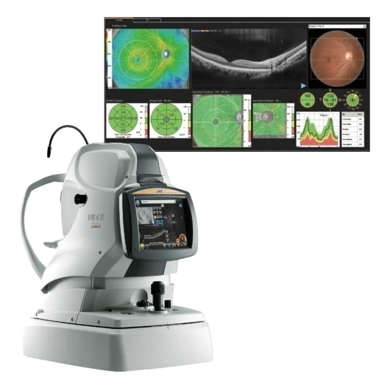
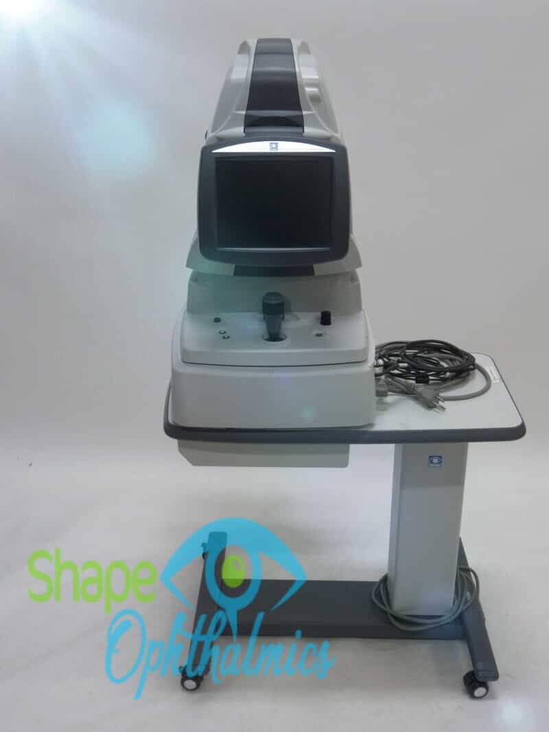
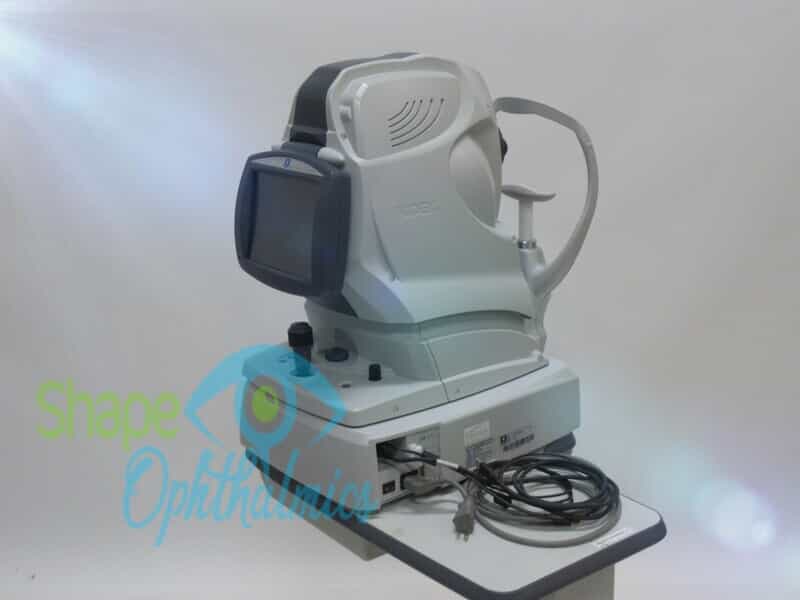
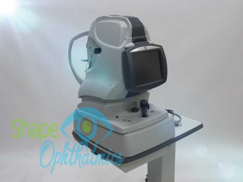
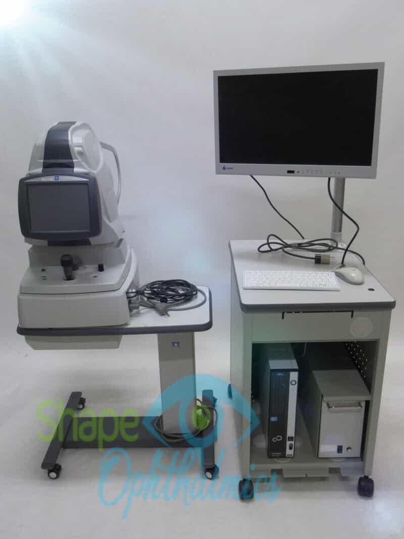
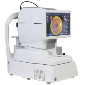
The Retina Scan Duo 2 is a combined OCT and fundus camera system that incorporates new features that enhance screening and clinical efficiency, as well as user-friendly features carried over from the previous model.
The intuitive software, automated functions, rapid measurements, and high-quality images make the Retina Scan Duo™ a pleasure to operate, akin to a camera capturing many of the vivid landscapes you’ve experienced over your lifetime. The combination of features results in a better overall experience for our patients and us.
FEATURES:
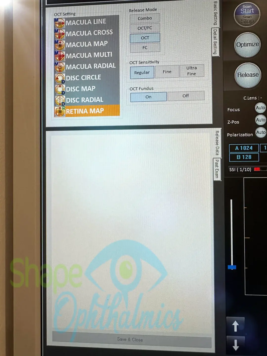
- Fundus image acquisition with macula and disc capture in one image on OCT
- Combined diagnosis of macular and disc pathologies
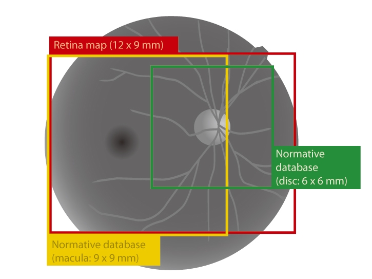
- Wide area scan (12 x 9 mm)
Wide area normative database (macula: 9 x 9 mm, disc: 6 x 6 mm): A 12 x 9 mm wide area image can be acquired. The retina map captures both the macula and disc in a single shot. The normative database provides a wide-area, color-coded map comparing the patient’s macular thickness to that of a population of normal eyes. - Denoising technique with deep learning: A new image enhancement method that automatically displays a denoised image once B-scan acquisition is complete. Using deep learning on a large dataset of images averaged from 120 images, this denoising technique provides high-definition images comparable to those from a multiple-image-averaging technique. The denoising function generates high-definition images from a single frame, reducing image acquisition time and increasing patient comfort.
- Quick acquisition of high-definition B-scan images from a single-frame image
- Fundus autofluorescence (FAF): The FAF function is an advanced screening feature that allows non-invasive evaluation of the RPE without contrast dye.
*Available for the FAF model
Combined diagnosis of macular and disc pathologies
Retina map
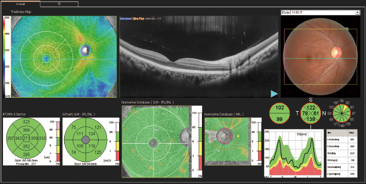
Wide area scan (12 x 9 mm)
Wide area normative database (macula: 9 x 9 mm, disc: 6 x 6 mm)
| A 12 × 9 mm wide-area image can be acquired. The retina map captures both the macula and disc in a single shot.
The normative database provides a wide-area, color-coded map comparing the patient’s macular thickness to that of a population of normal eyes. |
 |
Denoising using deep learning
A new image enhancement technique using deep learning automatically displays a denoised image once B-scan acquisition is complete. With deep learning of a large data set of images averaged from 120 images, this denoising technique provides high definition images comparable to a multiple-image-averaging technique. The denoising function generates high definition images from a single frame while decreasing image acquisition time and increasing patient comfort.
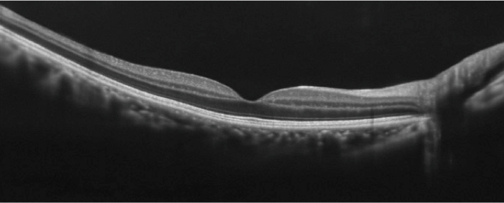
Denoised from a single-frame image |
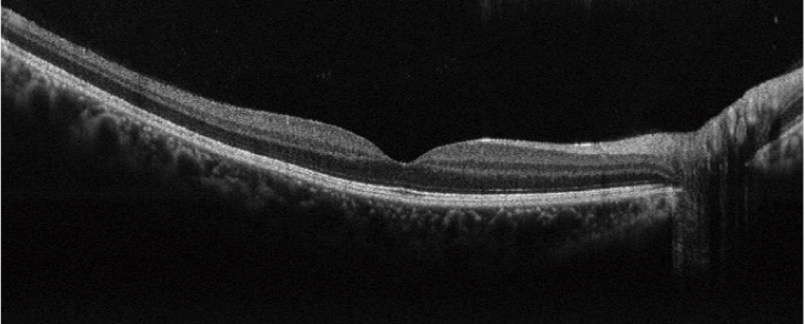
Averaged from 50 images |
Fundus autofluorescence (FAF)
| The FAF function is an advanced screening feature that enables noninvasive evaluation of the RPE without the use of contrast dye.
*Available for the FAF model |
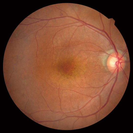
Color fundus image |
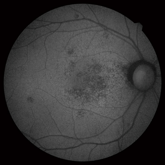
FAF image |
Optional features
AngioScan
| Details are available on the AngioScan page. | 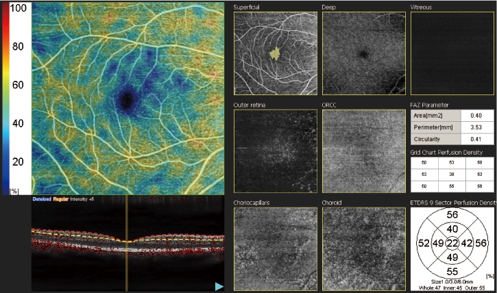 |
Long axial length normative database
Details are available on the long-axial-length normative database page.
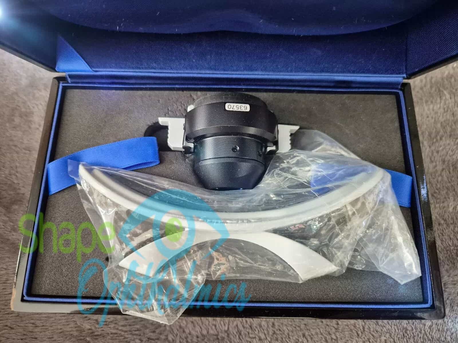
Anterior segment adapter
The optional anterior segment adapter enables observation and analysis of the anterior segment.
Reviews
There are no reviews yet.