Description
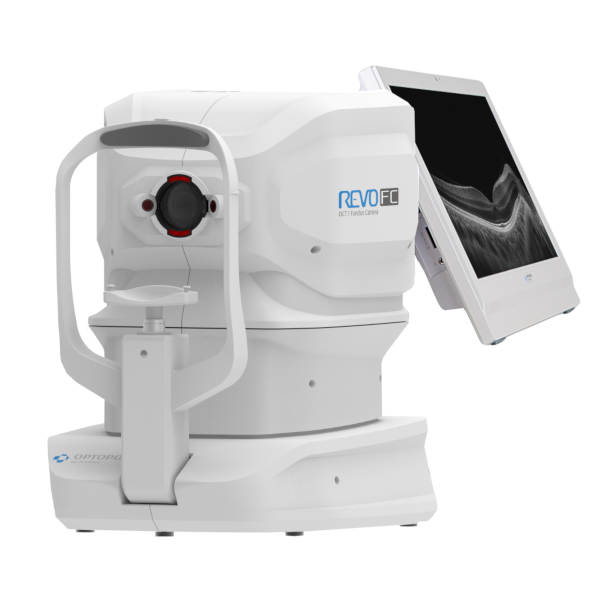
Optopol Revo FC offers all the proven advantages of REVO systems with state-of-the-art color imaging for a new level of diagnostic certainty. High-quality OCT scanning and comprehensive analysis of the retinal layers combined with fundus imaging make the examination more versatile than ever.
Optopol Revo FC is an OCT combined with Fundus Camera. A single versatile tool that combines high-resolution OCT images with true color background images. Acquisitions are fully automatic for Retina, Optical Disc, and Anterior Segment.
The tool can be implemented with the Angiography module (optional) and the Biometry module (optional). Like all Optopol OCT models, REVO FC interfaces with Optopol Perimeters for a simultaneous structural and functional damage analysis.
Optopol Revo FC Key Features:
- 80,000 scans/second
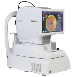
- True Color NonMyd. Integrated fundus camera
- OCT-A: OCT angiography
- B-OCT: Biometrics module
- Anterior Wide Field Adapter
- Very compact and fits in every room
- Retina – fundus camera – glaucoma – anterior segment
- Fully automatic one-touch measurement
- Fully equipped, good price
Optopol Revo FC offers all the proven benefits of REVO systems with state-of-the-art Fundus imaging technology for a new diagnostic confidence level. High-quality OCT scanning and comprehensive retinal layer analysis combined with Fundus imaging make examination versatile like never before.
What makes the REVO FC truly unique is the integrated non-mydriatic 12.3 Mpix Fundus camera capable of capturing extremely high-quality and detailed color images. The REVO FC Fundus Camera is fully automated, safe, and easy to use.
Optopol Revo FC, with a scanning speed of 80,000 A-scans / sec, offers advanced technologies and remarkable simplicity of operation. Meets all requirements for modern optical tomography.
The Copernicus REVO FC offers:
 3D retina
- Up to 12 X 12 mm scan area
- 8 layers are recognized automatically
- Automatic follow-up
- Normative Databases for Retinal Thickness and Ganglion Complex
- Progress display of max. 6 examinations
- Comparison of any two studies
 fundus camera
- True color fundus camera
- 45° Non-Mydriatic
- 12 megapixel CCD camera
- Fully automatic photography
 Real-time hardware eye tracking
- Active tracking in real time
- Perfect help with eye movement and blinking
- Accurate OCT images
- For precise follow-up examinations
 glaucoma
- Geometrical evaluation of the optic nerve (cup/disc)
- Nerve Fiber Layer Analysis (GDX)
- DDLS analysis
- Ganglion Complex Analysis
- Automatic follow up
- Normative databases for geometries (cup/disc), nerve fibers, ganglion cells
- Progress display of up to 6 examinations
 ganglion cell analysis
The REVO OCT’s ganglion cell analysis provides an additional glaucoma analysis to the conventional papillary retinal nerve fiber layer thickness measurement. You can choose between three analysis forms: NFL+GCL+ IPL Thickness, GCL+ IPL Thickness and NFL Thickness, additionally supported by respective NDBs.
Combining ganglion cell analysis with papillary retinal nerve fiber layer thickness provides a better biomarker for managing glaucoma.
 Anterior wide field
- Integrated front section module (5 x 5 mm)
- Automatic pachymetry
- Analysis of the chamber angle
Optional wide-angle module (14 x 5 mm):
- Representation of the complete front system
- Measurement of two chamber angles in one exposure
- representation of the iris
 OCT biometrics
Fully automatic 10-fold measurement:
- AL: axis length
- ACD: anterior chamber depth
- LT: lens thickness
- CCT: corneal thickness
In addition, there is a scanning program for a detailed display of the anterior chamber. Further information on the REVO Angio Module can be found in the module description.
 OCT topography
The OCT Topography (OCT-T) module enables a comprehensive analysis of the two interfaces of the cornea. The most important analyzes include determining the radii of curvature (K1 and K2) of the cornea and the resulting refractive power of the cornea.
The values complete the OCT Biometrics Module (OCT-B) of the Copernicus REVO series released in version 8.0. A large number of different representations are available for evaluating the results. The OCT-T technology is based on the principle of ray tracing. In addition to the front and back surface of the cornea, the corresponding corneal thickness is also determined.
Connectivity / Network
- Free server and viewer programs (optional installation service)
- Viewers may be redistributed to affiliated stores/locations
- site networking
- Various interfaces to your management software
- DICOM
- Automatic export
Optopol Revo FC Technical specifications:
- Technology: spectral domain OCT
- Measurement modes: B-scan, radial scan, grid, cross, 3D, grid scan, multiline scan, vessel scan Measurement
- method: fully automatic
- Fundus display: pSLO (live fundus reconstruction)
- Fixation: OLED screen (fovea and disk) – variable position and shape, external fixation
- Wavelength: 840nm
- Axial resolution: 5 microns
- horiz. Resolution: 12-18 microns
- Axial scan depth: min. 2mm
- Speed: 80,000 ascans/s
- Bscan width: up to 12mm
- Max. area 3D scan: 12 X 12mm
- max. A-scan / B-scan: 10,500
- Min. pupil: 2mm Device dimensions : 47.9 x 49.3 x 36.7 (WxHxD)
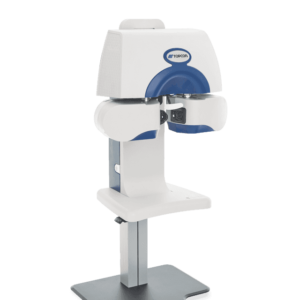
- Dimensions with table (STM-2): 88 x 129-154 x 53cm (WxHxD)
analysis software
- Retina Analysis:
Retina Thickness
RNFL+ GCL+IPL Thickness (Nervear Fiber Layer, Ganglion Cell Layer, Inner Plexiform Layer Thickness)
GCL+IPL Thickness
RNFL Thickness
RPE Deformation Map
IS/OS Thickness
Follow Up - Glaucoma:
Nerve Fiber Layer Thickness
ONH Morphology
DDLS – Disc Damage Likelihood Scale Ganglion Cell Analysis
with Asymmetry Comparison
Follow Up - Anterior:
Pachymetry
LASIK flap measurement
Chamber angle measurement
Wide-angle scans (with optional wide-angle module)
Include:
- Copernicus REVO NX FC OCT
- Anterior Module Standard (5mm)
- Viewer and Server applications
- HP All-In-One professional PC with 24″ multi-touch display table package (equipment table, PC/monitor holder, medical isolating transformer)
- OCT-A: OCT angiography plus
- T-OCT: topography module
- B-OCT: biometry Module
- anterior wide-field adapter
The Optopol Revo FC OCT Fundus Camera uses the latest OCT technology to capture cross-sectional retina images with exceptional detail and accuracy. Its advanced software and processing capabilities allow us to analyze these images in real time, providing valuable information about the thickness and health of the retina and other structures.
In addition, the system also includes fundus imaging capabilities, allowing us to capture high-quality images of the retina and other structures. These images can be used for diagnosis, monitoring, and patient education, making it an invaluable tool for ophthalmologists.
The Optopol Revo FC, OCT Fundus Camera, also offers advanced features, such as automated segmentation and layer analysis, which help to enhance further the accuracy and speed of diagnosis and monitoring of ocular conditions.
In conclusion, the Optopol Revo FC OCT Fundus Camera is an excellent choice for ophthalmologists who want to provide the best possible care for their patients. Its advanced imaging capabilities and precise measurements make it a valuable tool for diagnosing and managing various ocular conditions.
Reviews
There are no reviews yet.