
Description
Xephilio OCT-A1 achieves both high-speed OCTA image generation and high image quality by using image processing technology using deep learning technology and a high-performance GPU (image processing semiconductor). Under the new brand name, “Xephilio, will be widely deployed in ophthalmic medical institutions.
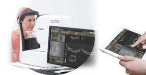
The Canon OCT A1, due to its innovative features, allows you to capture easily and fast with an incredible level of detail, fully automated with only three clicks. Thanks to its fully customizable settings and Ultra high definition OCT images, it is easy to use for all types of users. For greater precision, up to 200 images are considered to deliver the best possible definition in each measurement. Thus the structure of the retinal layers can be observed with the highest level of detail.
Xephilio OCT A1 features are:
- A single scan offers images with minimized noise and a high level of detail, allowing a comfortable examination for the patient.
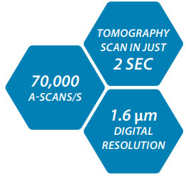
- It has the excellence of Canon optics that offers an image with a high resolution of 1.6um and a high scanning speed of 70,000 a-scans/s in 2 seconds.
- Intelligent functions that allow automated exams.
- Flexible information possibilities and extensive normative databases: It has extensive DICOM and EMR capability
- Anterior segment analysis: Available with the ASA-1 adapter that allows quick and easy quantification of standard parameters.
- OCT angiography: OCT angiography is a sophisticated technology that detects the movement of red blood cells in the retinal vasculature and allows the entire vascular network to be visualized in detail.
Improved OCTA clinical results thanks to the combination of the intelligent Denoise systems and the sophisticated Flow fusion technology. - Angio HD optional software – Delivers images with higher pixel density and an expanded field of view without losing image resolution, even from wide angles. And it also gives you the option to choose between square and rectangular formats up to 696 x 696 pixels.
- Retinal Expert software: protects the privacy of your patients and allows you to document your studies properly
Drawing on Canon’s renowned optical expertise, the Xephilio OCT-A1 delivers excellent image quality. With a digital resolution of up to 1.6 μm, the system enables excellent differentiation of individual retinal structures and layers. The high scanning speed of 70,000 A-scans/s allows for concise exam times, typically around two seconds, resulting in fewer motion artifacts and greater patient comfort.
FAST, EASY ACQUISITION WITH INCREDIBLE DETAIL
For outstanding performance and exceptional ease of use you can rely on every day, look no further than Xephilio OCT-A1. Superior image quality and various automated features optimize and simplify your examinations. At the same time, the system’s high scanning speed enables short examination times, increasing your efficiency and patients’ comfort.
THE NEW RETINAL IMAGING REALITY
OUTSTANDING IMAGING IS YOUR BEST FRIEND
Accurate scanning, outstanding ease of use

The system’s integrated Scanning Laser Ophthalmoscope (SLO) contributes significantly to scan quality and ease of use. Providing real-time retinal tracking enables accurate monitoring of the examination.
Fast and precise follow-up

The SLO also enables accurate follow-up examinations by automatically adjusting to the same scan position used in the previous exam. For reliable comparison, the software automatically selects identical scan parameters.
ACHIEVE CONSISTENTLY HIGH IMAGE QUALITY, AUTOMATICALLY
At times, involuntary eye movement during examinations is unavoidable. With its integrated SLO real-time retinal tracking technology, the system allows you to maintain the same scanning position automatically. As a result, Xephilio OCT-A1 retinal tracking significantly reduces movement artifacts and thus provides consistent, high image quality.
High definition, enhanced depth, wide field of view
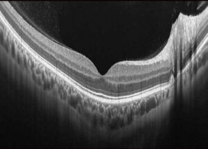
With Xephilio OCT-A1, you can average up to 200 scans to achieve an image resolution that allows you to see both the layer structure and the vitreous pleated structure in detail. For optimal imaging, the system offers unique scan modes for vitreous and choroid imaging, as well as a vast scan width of up to 13 mm
Reliable 10-layer recognition
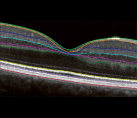
Thanks to its excellent image quality and resolution, Canon’s Xephilio OCT-A1 can automatically detect and distinguish ten retina layers – including Bruch’s membrane (BM).
FAST AND EASY ACQUISITION WITH INCREDIBLE DETAIL
Examinations with the Xephilio OCT-A1 are straightforward to delegate. A complete range of intelligent functions enables fully automated examinations. The auto-re-scan function intervenes if a patient makes unwanted eye movements and automatically compensates for any artifacts.
Automated anterior tracking
After pointing approximately to the center of the pupil, the system automatically detects and maintains the exact center, even when the patient is moving his eye or blinking.
Automatic image optimization
Afterward, the built-in autofocus and C-gate functions will automatically determine the highest signal quality for the best possible examination results.
Real-time retinal tracking
By detecting and compensating movements in the fundus images on a frame-by-frame basis, the impact of small involuntary movements is reduced during fixation, and motion artifacts are significantly reduced.
FAST, CONSISTENT EXAMS – HIGH PATIENT COMFORT
Xephilio OCT-A1 offers ten fixed, freely programmable exam presets, allowing you to combine multiple scan modes into a single exam. Using presets can help you improve the workflow and consistency of exams and, at the same time, increase patient comfort.
Versatile reporting possibilities, extensive normative databases
Xephilio OCT-A1 provides a full range of reporting tools, including relevant normative databases. Thanks to its extensive DICOM and EMR capability, results from multiple Canon imaging modalities can be stored, shared, and analyzed as needed in your daily practice.
Macula
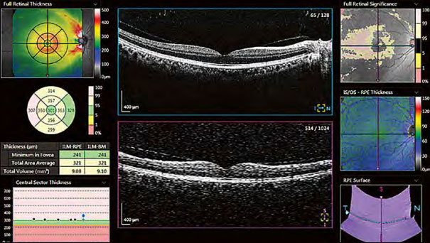
The system provides a detailed retinal thickness analysis using comparisons with normative databases, ETDRS grids, tables, and 3D visualizations.
Glaucoma
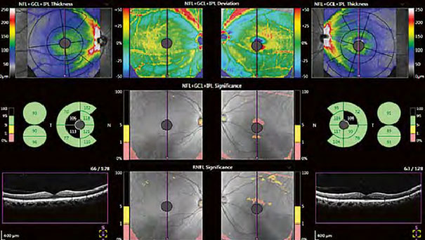
Early detection is the key to slowing the progression of glaucoma. Xephilio OCT-A1 supports NFL + GCL + IPL and GCL + IPL measurements with a comprehensive set of graphical representations for complete analysis.
COMBINED REPORT
By sharing the same database with an optional retinal camera, fundus images can be easily integrated into the OCT evaluation. Fundus and OCT images can be displayed side by side, mapped, and superimposed as needed.
Optic Disc
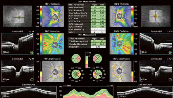
Xephilio OCT-A1 allows a comprehensive analysis of all optic disc parameters, including comparisons with an extensive normative database.
Progression report
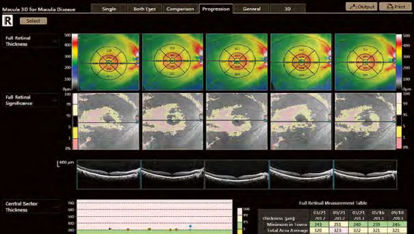
Analysis results comparing five examinations arranged in the time sequence of eyes on the same side in the same scan mode and the same size of the scanning area.
ANTERIOR SEGMENT ANALYSIS
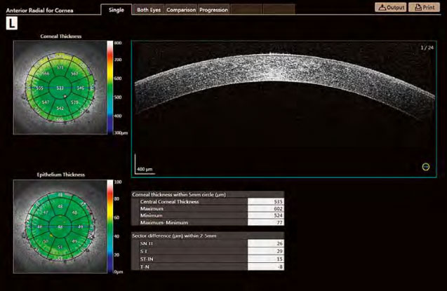
With the optional Anterior Segment Adapter ASA-1, Xephilio OCT-A1 also allows you to analyze and document the anterior segment of the eye during the same exam. The included measurement package will enable you to quantify standard parameters quickly and easily.
The corneal thickness analysis on Xephilio OCT-A1 is presented as corneal and epithelium thickness maps, including corneal grids and a numerical table.
The anterior segment analysis kit allows you to measure the distance between two points, arbitrary angles, as well as AOD (Angle Opening distance) and TISA (Trabecular Iris Space Area) values.
In addition, the system includes a range of imaging modalities and capture modes, allowing us to tailor the imaging process to each patient’s specific needs. Its advanced software also offers a variety of analysis tools, making it easy to interpret the data and diagnose ocular conditions.
The Canon Xephilio OCT-A1 also offers a range of advanced features, such as automated segmentation and layer analysis, which help to enhance further the accuracy and speed of diagnosis and monitoring of ocular conditions.
In conclusion, the Canon Xephilio OCT-A1 is an excellent choice for ophthalmologists who want to provide the best possible care for their patients. Its advanced imaging capabilities and precise measurements make it a valuable tool for diagnosing and managing various ocular conditions.
Source
Reviews
There are no reviews yet.