Description
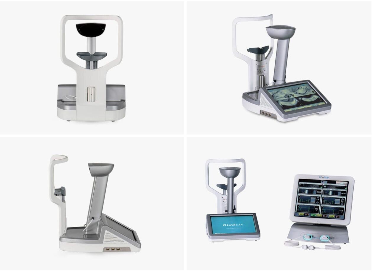
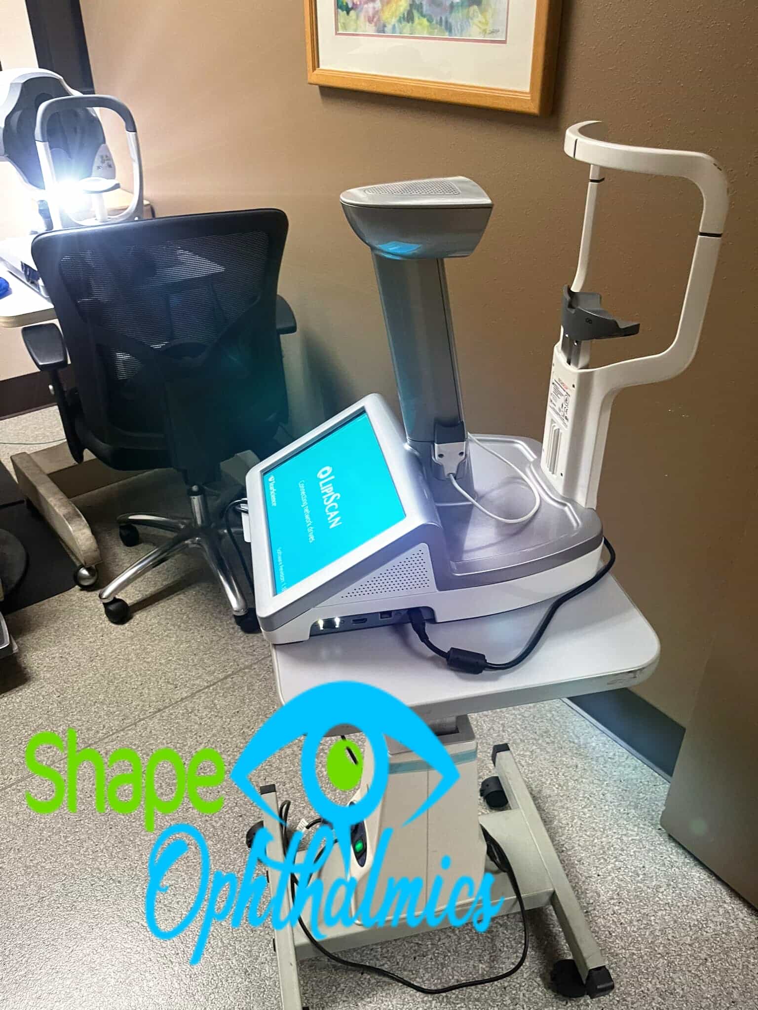
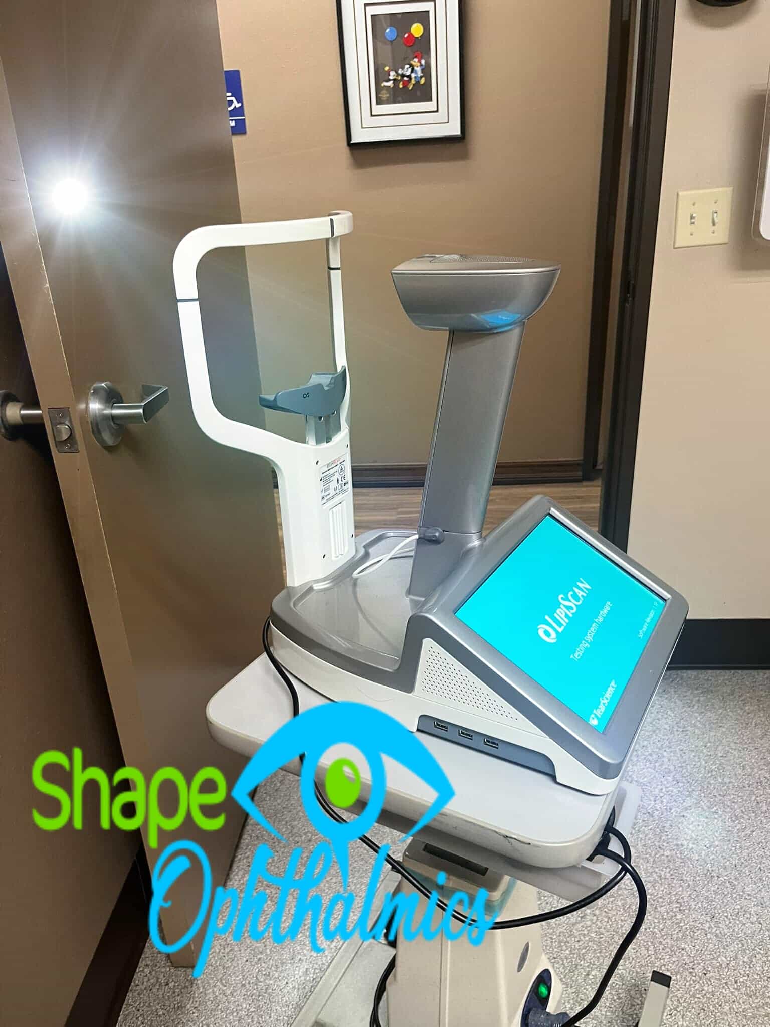
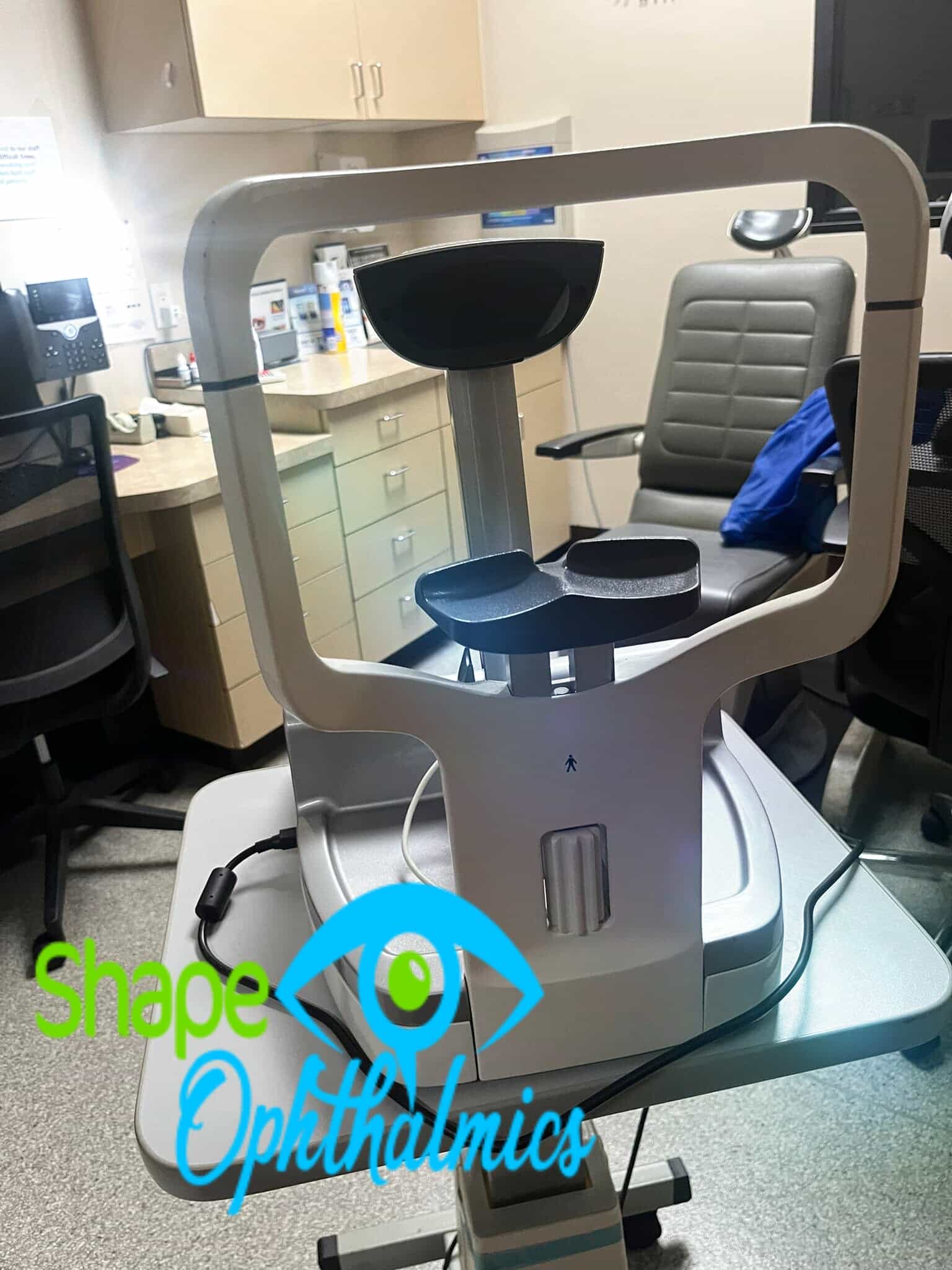
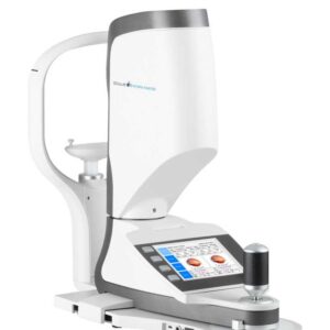
The TearScience LipiScan Dynamic Meibomian Imager (DMI) is a special eye device. It helps to see and check the meibomian glands in the eyelids. This tool is essential for diagnosing meibomian gland dysfunction (MGD). MGD is a significant contributor to dry eye disease.
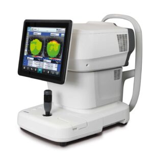
A dedicated HD meibomian gland imager designed for efficiency and versatility.
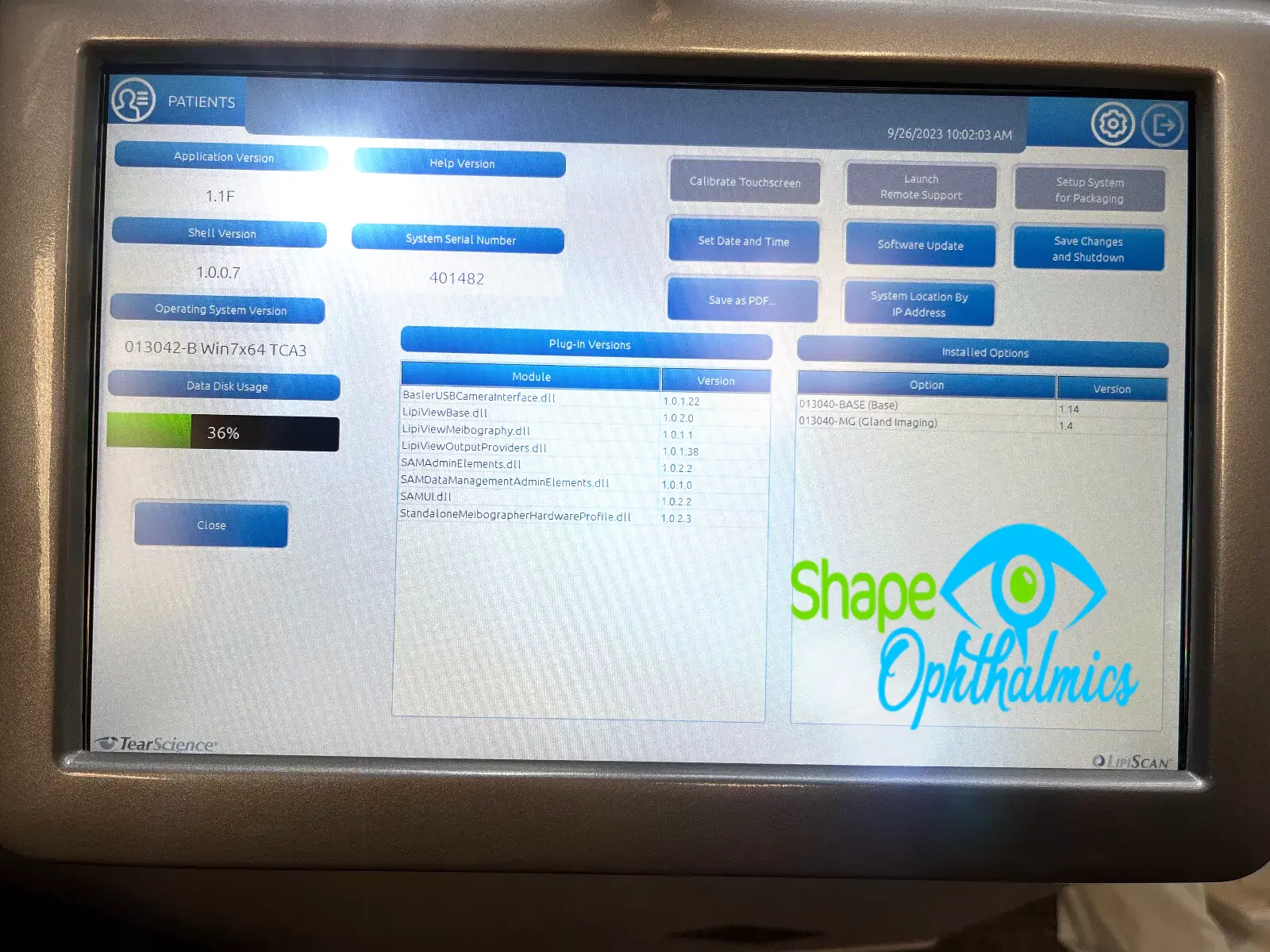
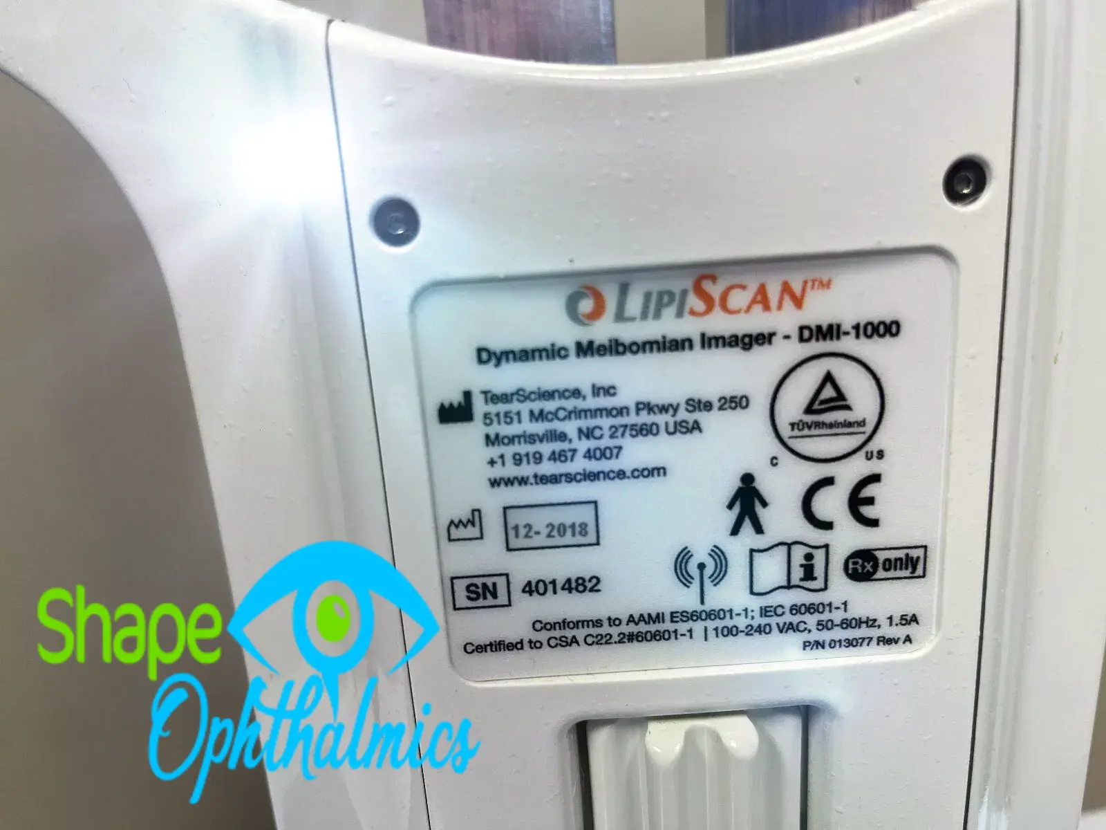
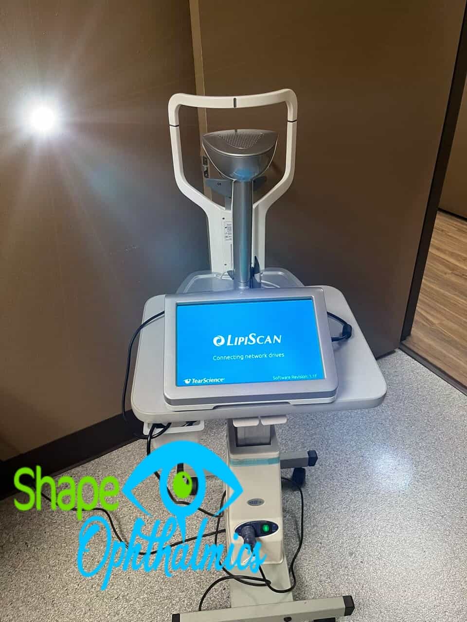
The TearScience LipiScan with Dynamic Meibomian Imaging (DMI) is a compact and user-friendly device. It makes high-definition meibography available for any practice.
The designers created the durable yet lightweight device to maximize workflow and enable easy integration into busy practices. Both lower eyelids can be imaged in about a minute.
Key Highlights
- High‑Definition Imaging
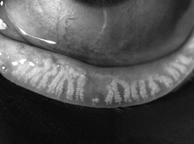
Uses near-infrared (NIR) light with a high-definition camera. This captures clear black-and-white images of the meibomian glands.
- Dynamic Illumination & Adaptive Transillumination
These patented techniques lower surface glare. They also adjust light intensity based on eyelid thickness. This leads to clearer and more detailed images.
- Efficient Workflow & Portability
Lightweight (approximately 11 kg or 25 lbs), compact, and designed for tabletop use. You can image both lower eyelids in about a minute. This makes it a great fit for clinical settings.
- Clinical and Regulatory Credentials
Carrying FDA 510(k) clearance (K182506), it’s classified as a Class II ophthalmic camera intended for adults. It captures and stores images of digital meibomian glands. However, it does not make diagnoses.
- Practical Features
The product has easy touchscreen controls. It offers simple export options, including PDF and DICOM, for electronic medical records (EMRs). It also meets medical-grade safety and disinfection standards from
Dynamic Meibomian Imaging
MEIBOMIAN GLANDS IN HIGH DEFINITION
Dynamic Illumination
Surface lighting originates from multiple light sources to minimize reflection.
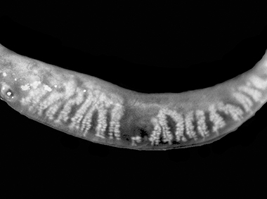
Adaptive Transillumination
Changes to the light intensity across the surface of the illuminator compensate for variations in lid thickness between patients.
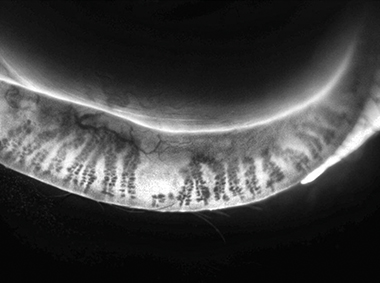
Dual-Mode DMI
A combination of Dynamic Illumination and adaptive transillumination provides an enhanced view of meibomian gland structure.
How TearScience™ LipiScan™ Works1
- Workflow maximization with fast capture of meibomian gland images
- Small footprint and lightweight (10 kg) for optimal versatility
- Fast and intuitive operation for seamless integration into routine workups
- Renders a high-definition image of the meibomian gland structure
- Option to export images in various formats
Why It Matters
- Improved Diagnosis: Enables clinicians to quickly evaluate gland structure and detect dropout, atrophy, obstructions, or morphological changes.
- Optimizes Dry Eye Management: Supports early diagnosis and helps monitor progress with treatments like LipiFlow® or eyelid hygiene routines.
- Easy Integration: Designed for busy clinics—fast operation, compact size, and compatibility with existing systems.
Who Should Use It?
- Ophthalmologists and optometrists assessing dry eye, eyelid disorders, contact lens tolerance, or pre/post-surgical surface health.
- Practices aiming for objective documentation and monitoring of meibomian gland structure over time.
The TearScience LipiScan Dynamic Meibomian Imager is an easy-to-use tool for doctors. It is FDA-cleared and provides quick, high-quality images of meibomian glands. Valuable for diagnosing and monitoring MGD, advanced imaging improves patient care by integrating into everyday eye exams.
Reviews
There are no reviews yet.