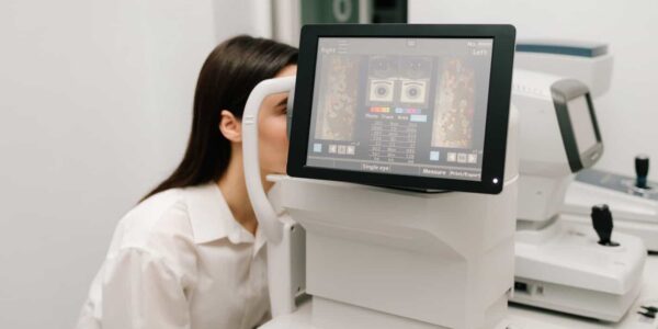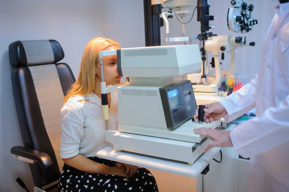
Fundus cameras are typically found in an ophthalmologist’s or optometrist’s office, and the procedure is usually done, outpatient. The patient will be seated in front of the camera, and the eye will be numbed with eye drops to make the procedure more comfortable. The eye will be held open with a speculum, and the camera will be positioned over the eye to take the picture.
A fundus camera is a specialized device that photographs the eye’s interior, specifically the retina and choroid. This medical imaging equipment typically includes a technical camera, flash, illuminator, and filters. It is invaluable for diagnosing and monitoring eye diseases such as glaucoma, macular degeneration, diabetic retinopathy, and more.
What is a Fundus Camera
A fundus camera is a medical device that takes images of the back of the eye, also known as the retina. It is an important tool for ophthalmologists and optometrists to diagnose and monitor various eye conditions, including diabetic retinopathy, glaucoma, and age-related macular degeneration. Ophthalmologists and optometrists use these images to detect and diagnose eye disorders, making fundus cameras a vital tool in eye care.
Several fundus cameras from Shape Ophthalmics are available today, each with features and capabilities. Some of the most common types of fundus cameras include:
- Non-mydriatic fundus cameras: These cameras do not require dilating drops, making them ideal for patients sensitive to these medications.
- Mydriatic fundus cameras: These require dilating drops, which allows the capture of more detailed retina images.
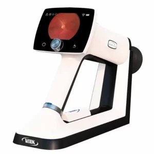
- Handheld fundus cameras are small and portable, making them ideal for use in remote or underserved areas.
- Fundus cameras with built-in imaging software: These cameras include software that enables immediate analysis of captured images.
Fundus camera imaging techniques
A fundus camera can use several imaging techniques to capture images of the retina. These include:
- Color fundus photography: This technique uses a flash of light and a color filter to capture a detailed retina image. This is the most common fundus imaging type used for routine screenings and diagnosis of retinal diseases.
- Fluorescein angiography: This technique involves injecting a dye into the patient’s bloodstream and capturing retina images as the dye flows through the blood vessels. This technique detects and diagnoses retinal diseases such as diabetic retinopathy and age-related macular degeneration.
- Indocyanine green angiography: This technique is similar to fluorescein angiography but uses a different dye to better visualize the choroid, a layer of blood vessels between the retina and the sclera. It’s also used to detect and diagnose retinal diseases.
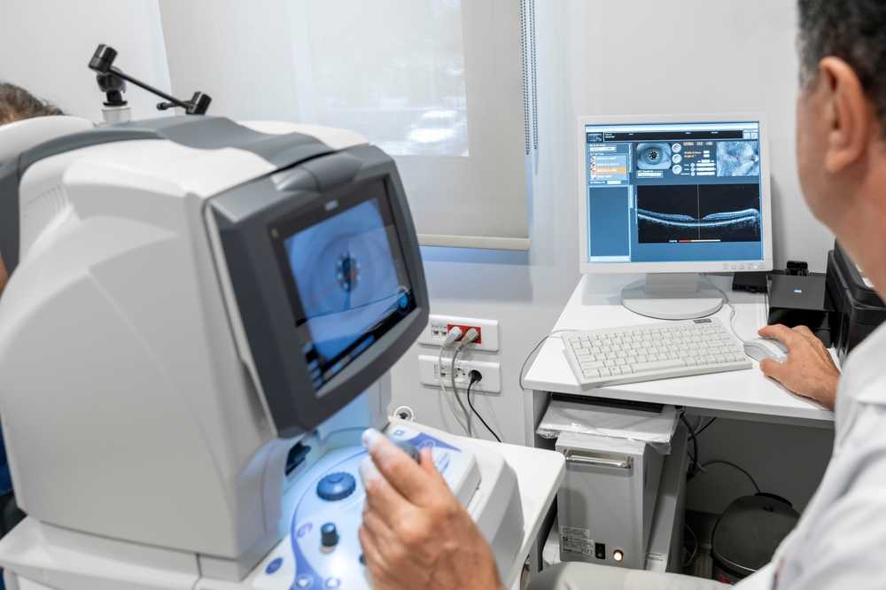
- Optical coherence tomography (OCT): This technique uses infrared light to capture detailed cross-sectional images of the retina. It helps detect and monitor the progression of retinal diseases such as macular edema and glaucoma.
- Fundus autofluorescence (FAF): This technique captures retina images using a special filter that detects the natural fluorescence of retinal pigments. It’s used to detect and monitor the progression of retinal diseases, such as age-related macular degeneration.
- Confocal scanning laser ophthalmoscopy (CSLO) uses a laser beam to scan the retina and capture high-resolution images. It detects and diagnoses retinal diseases such as age-related macular degeneration, diabetic retinopathy, and retinal detachment.
These are some examples of the most common fundus camera imaging techniques, but new technologies and methods are constantly being developed.
What are the Benefits of Fundus Cameras
The retina is part of the eye responsible for capturing light and sending visual signals to the brain. It comprises several layers of nerve cells, including the photoreceptors that detect light and the ganglion cells that transmit visual information to the brain. The retina also contains blood vessels that provide oxygen and nutrients to the eye.
A damaged or diseased retina can cause many symptoms, including blurred vision, blind spots, and even blindness. A fundus camera can help ophthalmologists and optometrists detect these problems early before they cause significant vision loss by taking pictures of the retina.
Fundus cameras work by shining a bright light into the eye and using a special lens to capture a high-resolution retina image. The camera can capture both color and black-and-white images, which can be used to detect various eye conditions. For example, diabetic retinopathy, which is caused by damage to the blood vessels in the retina, can be identified by changes in the color of the blood vessels.
Here are some benefits of fundus cameras for the patient, ophthalmologists, and optometrists.
- Improved diagnosis and treatment: Fundus cameras provide ophthalmologists and optometrists with detailed and accurate retina images, which helps them identify and diagnose eye disorders early on.
- Preservation of vision: Fundus cameras help to detect and monitor eye diseases early on, which is crucial in preserving vision.
- Better patient outcomes: By using fundus cameras, ophthalmologists and optometrists can adjust treatment plans based on the progression of a patient’s condition, leading to better outcomes.
- Cost-effective: Fundus cameras are cost-effective as they are non-invasive and do not require any anesthesia.
- Easy to use: Fundus cameras are user-friendly, and results can be obtained instantly with built-in image analysis software.
- Widely available: Fundus cameras are widely available and can be found in most ophthalmology and optometry clinics.
Images taken with a fundus camera are also used to monitor the progression of eye conditions over time. For example, suppose a person has diabetic retinopathy. In that case, their ophthalmologist may take regular pictures of their retina to see how the condition progresses and decide on the treatment.
The fundus camera is a noninvasive, safe, and painless examination, making it a handy tool for patients who have difficulty with other eye examination methods, such as children, the elderly, or patients with neurological conditions.
Fundus Camera product in the market.
When it comes to the top and most wanted Fundus Camera products in the market, a few stand out from the rest.
Many models now include auto-focus capabilities, digital zoom capabilities, up to 8x magnification, and enhanced lighting systems for improved image capture and analysis software, allowing physicians to quickly review images and make precise measurements or annotations directly on them. With such capabilities and increasing affordability and availability, fundus cameras have become an essential tool for ophthalmologists worldwide.
Here are some of the top and most wanted fundus camera products currently on the market:
1. iCare DRSplus ($6,999.00)

The iCare DRSplus is a revolutionary eye-care tool that provides exceptional imaging capabilities for detailed retina examination. This system provides clinicians with a wide field of view, high contrast and resolution, and color accuracy that can help detect various ophthalmic diseases. The combination of the TrueColor Confocal technology, superior optical performance, and intuitive software design enables faster diagnosis and improved patient care.
In addition to its impressive features for examining the retina itself, the iCare DRSplus TrueColor confocal fundus imaging system also boasts superior optics for assessing cataracts and other anterior segment characteristics as well, through its multiple imaging options, including:
- UHR OCT scans of both anterior segments and posterior segments
- 3D scanning capability; wide field digital photography
- anterior segment OCT scans
- enhanced list mode viewing
- nonmydriatic photography
- autofluorescence imaging, etc.,
2. Volk Pictor Prestige ($4,995.00)
The VOLK Pictor Prestige Portable Fundus Camera employs advanced technology to provide high-resolution and detailed retina images, making it an essential tool for diagnosing and treating glaucoma, macular degeneration, and diabetic retinopathy. It is ideal for ophthalmologists, optometrists, and other eye care specialists looking to improve diagnostic capabilities and streamline workflow.
3. Topcon NW500 ($18,900.00)
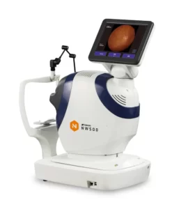
The Topcon NW500 Non-mydriatic Retinal Camera’s advanced technology and user-friendly design make it ideal for capturing high-quality retina images. It is also an essential device for diagnosing and treating various eye conditions, such as glaucoma, macular degeneration, and diabetic retinopathy.
Overall, the Topcon NW500 Non-mydriatic Retinal Camera is an excellent investment for any eye care practice seeking to enhance its diagnostic capabilities and streamline its workflow. Its exceptional features, reliability, and user-friendly design make it a valuable asset that will improve patient outcomes and overall practice efficiency.
4. Zeiss Clarus 700 ($21,999.00)

The ZEISS Clarus 700 utilizes advanced optical biometry and digital imaging technology for fast, accurate measurements and diagnoses.
The Clarus 700 has a unique dual-camera system, with one camera dedicated to color photography and the other to infrared imaging. This allows the system to capture widefield color and multicolor IR images simultaneously at different wavelengths, providing higher-resolution imaging than previously possible with traditional single-camera methods.
The Clarus 700 is also equipped with Zeiss Flourescite technology, which uses the fluorescence of certain dyes injected into the eye to highlight blood vessels and diagnose diseases like diabetic retinopathy more accurately than conventional methods.
In summary, ZEISS Clarus 700 is an ideal choice for busy clinical practices, helping maximize efficiency in the clinical setting.
5. Optomed Smartscope Pro Handheld Fundus Camera ($3,900.00)
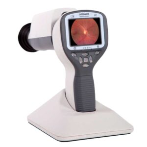
The Optomed Smartscope Pro Handheld Fundus Camera captures high-quality images of the retina and other ocular structures. This portable, lightweight device offers advanced features and an intuitive design, making it an excellent choice for ophthalmologists in various settings.
Optomed Smartscope Pro Handheld Fundus Camera is an excellent choice for ophthalmologists who want to provide the best possible care for their patients. Its advanced features, ease of use, and portability make it a versatile and valuable tool for capturing high-quality images of the retina and other ocular structures.
Conclusion
In conclusion, a fundus camera is vital for detecting and monitoring eye conditions, including diabetic retinopathy, glaucoma, and age-related macular degeneration. By taking high-resolution retina images, ophthalmologists and optometrists can detect problems early before they cause significant vision loss. The procedure is non-invasive, safe, and painless, making it an accessible examination for many patients.
Suppose you have been experiencing any symptoms of eye problems. In that case, it is important to schedule an appointment with an eye specialist who can use a fundus camera to examine your retina and provide an appropriate diagnosis and treatment.
It’s worth noting that the ranking of these products may change over time as new products are introduced to the market and existing products are updated. Also, different users may have different preferences and requirements for their fundus cameras, so always consult a professional or conduct further research before purchasing.