One of the primary goals of routine eye exams is to detect signs of eye disease before they cause vision changes or vision loss. The moment that a provider notices early signs of glaucoma or diabetic retinopathy is a pivotal moment in patients’ lives because identifying the disease means they can begin treatment that will prevent complications in the future.
Traditional diagnostic tools improve early detection rates, but they have drawbacks. These systems offer limited retinal visibility, potentially leading to missed peripheral pathology. The latest generations of retinal imaging devices integrate digital imaging, offering greater clinical delta than manual observation. Today’s retinal scanner machines range from compact fundus cameras to advanced ultra-widefield systems — each built to catch what older tools miss.
Ultra-widefield imaging tools capture high-resolution images that show more of the retina than traditional methods do, enabling a more complete evaluation of eye health. Learn how Ultra Widefield Imaging can improve early detection and how to choose between top machines to find the best fit for your practice.
What Is Retinal Imaging?
Retinal imaging is a standard part of routine eye care. Visualizing the retina allows providers to identify structural changes associated with retinal disease. This provides the opportunity to treat eye disease before patients become symptomatic.
How It Works
Retinal imaging is a non-invasive screening process that captures images of the retina, optic nerve, and blood vessels. The images are used for diagnosis, monitoring, and documentation of eye conditions.
Why It Matters in Daily Practice
Providers use retinal imaging to evaluate the overall health of the eye. The clinical information gained from retinal images allows providers to detect common retinal pathologies such as:
- Diabetic retinopathy
- Macular degeneration
- Glaucoma
Clinicians can use the images produced to help explain findings to patients. Patients can see evidence of changes to their eyes, which helps them understand a diagnosis. Sequential imaging can be a tool for showing patients changes over time and demonstrating how treatment adherence improves outcomes.
Imaging can also reduce referrals for complex treatments by stabilizing eye health through early intervention.
Common Types of Retinal Imaging
There are a variety of retinal imaging technologies available for eye care practices. The practice’s size, location, and patient base influence which type of retinal imaging device clinicians choose.
Fundus photography: A specialized camera captures high-resolution images of the retina, macula, and optic nerve. Both table-top and portable devices are available.
Optical Coherence Tomography (OCT): OCT is a cross-sectional imaging technology that creates detailed 3D images of retinal layers. It measures retinal thickness, which is useful for diagnosing conditions such as glaucoma and macular degeneration.
Fluorescein Angiography (FA): Fluorescein angiography is a technique that involves injecting a specialized fluorescent dye into the patient’s veins. The dye highlights the eye’s vascular structures, allowing eye doctors to detect leaks, blockages, or other problems in the retinal vessels.
Ultra-widefield (UWF) Imaging: Captures high-resolution images of the retina. Unlike traditional imaging, UWF can capture up to 200°, or 82% of the retina in a single scan.
While imaging systems all perform the same essential function, not all imaging is equal. Some systems see far more than others.
Ultra Widefield Imaging — Seeing Beyond the Central Retina
Ultra-widefield retinal imaging provides clinicians with a broader view of the retina than traditional cameras do. It increases the visibility of peripheral pathology, improving detection of detachments, tumors, and diabetic retinopathy.
How It Works
UWF cameras use laser-scanning technology to capture up to 200° of the retina, or 82%. This is a significant improvement on the 45° view that traditional cameras can capture. These devices often don’t require pupil dilation, so patients don’t have to cope with blurring and light sensitivity while they wait for the duration drops to wear off.
Step-by-Step Workflow in a Clinic Patient
Capturing images with UWF retinal imaging is quick and simple. Since it doesn’t require dilation, there is no lag time for patients waiting for the drops to take effect. This improves workflows and allows practices to increase throughput.
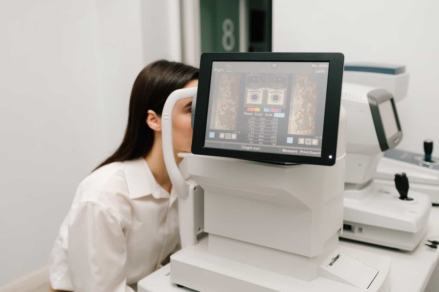
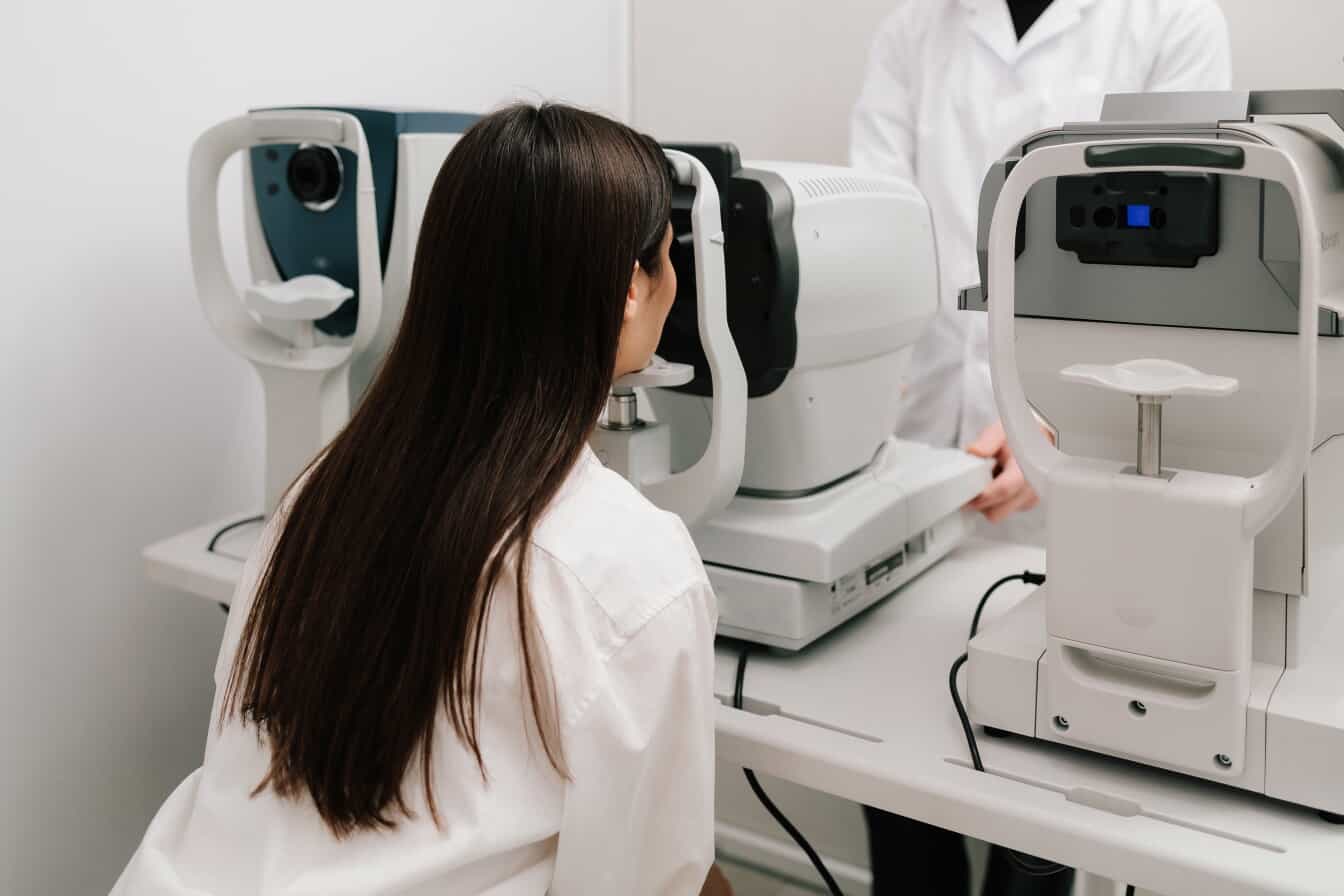
- Positioning: Patients sit in front of the machine, place their chin on a rest, and rest their forehead against a bar.
- Fixation target: The staff member conducting the screening instructs patients to fixate on a specific point, usually marked by a green light.
- Rapid image capture: Once the patients’ eyes are properly aligned, the clinician initiates the scan. Patients see a quick flash of light as the lasers capture the retinal image. There is no contact or puff of light. The process is repeated with the second eye immediately after the first.
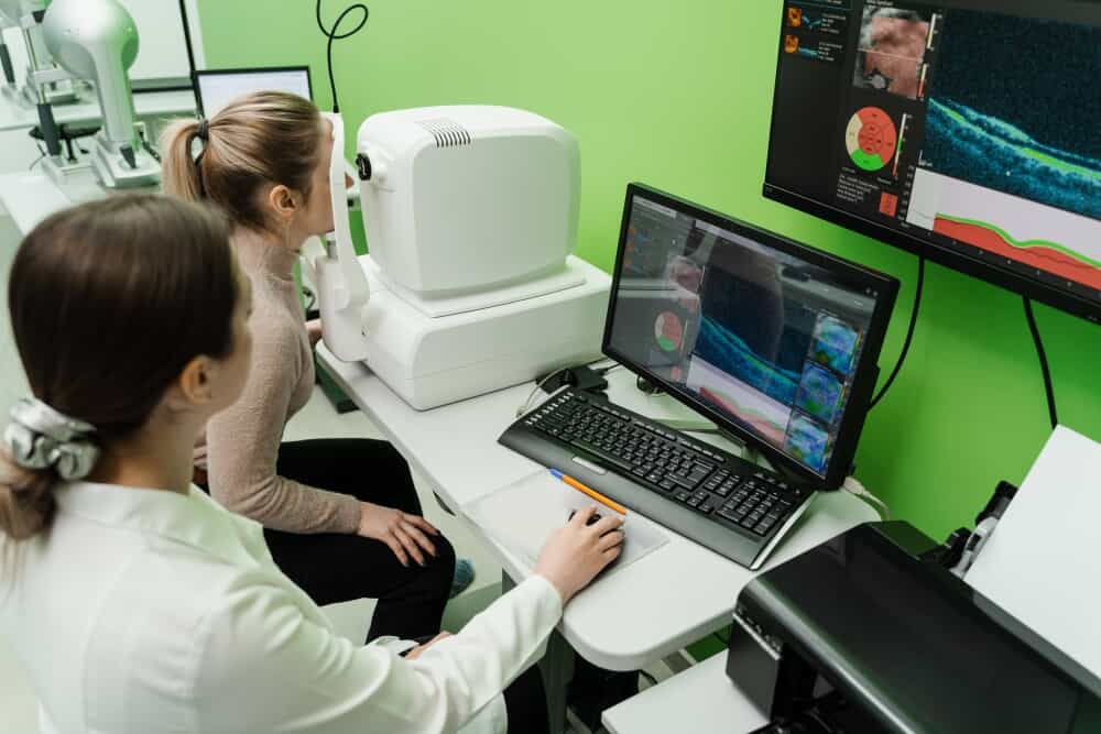
- Software processing: The UWF device automatically processes and uploads images, allowing eye care providers to review them immediately. Patients know their results during their appointment.
Why Clinics Are Switching
Eye care practices are upgrading to UWF devices for the clinical benefits they convey. The broader field of view of each retinal image enables providers to see more of the retina, facilitating earlier detection of peripheral lesions and other signs of eye disease.
The speed and ease of UWF scanning benefit patients, who appreciate not having to undergo pupil dilation. The detailed images provide high diagnostic confidence and better outcomes through earlier detection.
Trade-Offs to Consider
Some practices find the upfront cost to be an obstacle. The initial investment is higher than traditional retinal imaging equipment, but the improved workflow can lead to practice efficiencies and higher patient throughput.
In addition, clinicians need to budget time to learn how to assess the images. Once providers are used to the new process, they typically find the images more useful than the traditional methods.
Choosing the Right Retinal Imaging Machine (What Actually Matters)
Every practice has a unique set of needs. Practice managers should consider clinical factors, costs, patient base, and growth plans before committing to a retail imaging system.
1. Imaging Technology
There are benefits to using all available retinal imaging technologies. Fundus cameras are useful for practices that see patients at low risk for retinal disease. OCT systems offer more detailed imaging in a format that’s familiar to many clinicians. Ultra-widefield technology provides a more comprehensive clinical view of the retina, making it beneficial for screening patients with risk factors for retinal diseases, such as diabetes, a family history of macular degeneration, or glaucoma.
2. Field of View
Standard fundus photography and OCT systems are staple screening modalities in many clinics. While familiar and reliable, it offers a limited view of the retina, capturing only about 45% of its structure. UWF systems allow clinicians to see more of the retina and do a more thorough evaluation. The broader field of view is critical in identifying signs of disease in the peripheral regions of the eye.
3. Image Quality & Resolution
Image clarity is key to detecting subtle signs of retinal disease. True color technology available in UWF systems delivers higher contrast, enhancing diagnostic interpretation compared to traditional processed imaging.
4. Ease of Use & Automation
Retinal screenings should be as simple as possible for both patients and staff. Machines that offer automated functions such as autofocus, alignment, and capture can screen patients more quickly than systems with manual settings.
Automation also reduces the amount of training staff need to learn how to operate the system.
5. Patient Comfort
Pupil dilation is both time-consuming and inconvenient for patients. It adds to appointment duration, and the vision distortions and dilation last for several hours. Non-mydriatic systems speed up exams and allow patients to return to normal activities as soon as their appointment is over.
6. Software & Integration
Interoperability with existing technologies improves the ease of use of retinal imaging systems. Advanced retinal imaging systems integrate with existing EMR and cloud storage systems to automatically upload images to patient files. AI-powered tools can assist with image interpretation, supporting better clinical decision-making.
7. Budget & ROI
The upfront cost of retinal imaging systems can be significant, but it’s an investment in your practice. A more efficient system can increase the number of patients you can see in a week, thereby boosting revenue from volume. Better diagnostic capabilities also enable you to provide treatment and monitoring for patients who need them, adding another revenue stream to your practice.
Prices for retinal imaging machines vary widely by technology. Entry-level fundus cameras typically start in the low thousands, while mid-range OCT systems run $15,000–$30,000. Ultra-widefield systems like the Optos Daytona can exceed $50,000 at list price. Most vendors offer financing or demo programs — contact your rep for current pricing on retinal imaging machines.
Now that you understand the criteria to consider when choosing a retinal imaging system, let’s look at real machines that clinics are using.
Top 10 Retinal Imaging Machines for Your Practice (2026)
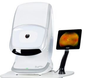
1. Optos Daytona ($8,888.00)
The Optos Daytona is an ultra-widefield system best suited for high-volume clinics. This non-mydriatic system captures up to 200° in under half a second per eye.
If you’re serious about improving retinal diagnostics and modernizing your workflow, the Optos Daytona is more than an upgrade—it’s a shift in how you deliver care.
It enhances efficiency, strengthens clinical confidence, and transforms patient communication.
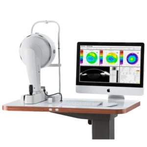
2. Oculus Pentacam ($11,450.00)
The Oculus Pentacam is a support system for capturing high-quality anterior imaging. It complements retinal diagnostics and is ideal for cornea-focused practices.
If you’re managing or growing an optometry or ophthalmology clinic, the Oculus Pentacam isn’t just another piece of equipment—it’s a strategic upgrade.
It simplifies your workflow, strengthens your diagnostics, and positions your clinic as a place where patients receive thorough, modern eye care.
And from one practice owner to another—that combination is hard to beat.
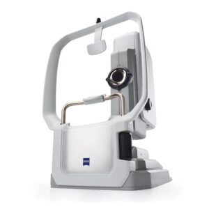
3. ZEISS CLARUS 700 ($14,999.00)
The ZEISS CLARUS 700 is a true-color widefield system. It’s ideal for practices that conduct advanced diagnostics.
If your goal is to elevate both diagnostic precision and patient communication, the ZEISS CLARUS 700 is a strong long-term investment.
It enhances how you diagnose, how you explain, and how your patients perceive the quality of your care.
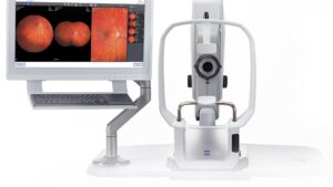
4. ZEISS CLARUS 500 ($9,999.00)
The ZEISS CLARUS 500 is a mid-range alternative to the ZEISS CLARUS 700. It boasts a compact design, making it a space-saving alternative to bulkier systems.
If you’re running or scaling an optometry or ophthalmology clinic, the ZEISS CLARUS 500 is more than just a retinal camera—it’s a complete imaging solution.
It improves how you diagnose, how efficiently your clinic runs, and how patients perceive the quality of your care.
5. Topcon Maestro2 ($15,999.00)
The Topcon Maestro2 is an OCT and fundus camera combination system. This robotic camera has advanced automation features for exceptional ease of use.
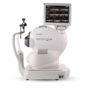
If you’re running a modern optometry or ophthalmology practice and want efficiency without losing diagnostic power, the Topcon Maestro2 is a smart investment.
It simplifies your workflow, standardizes your imaging, and helps you deliver consistent, high-quality care—day in and day out.
6. iCare DRSplus ($6,999.00)
The iCare DRSplus is a portable confocal fundus imaging system that provides high-quality TrueColor images. It features a user-friendly interface ideal for routine screenings.
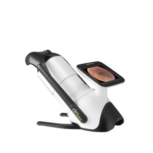
If you’re looking for a reliable, automated retinal imaging system that improves both workflow and patient comfort, the iCare DRSplus is a strong choice.
It doesn’t try to be everything—but what it does, it does efficiently and consistently.
7. Topcon NW500 ($18,900.00)
The Topcon NW500 is a fully automated non-mydriatic retinal camera system. This high-efficiency device is best for practices that need to optimize workflow.
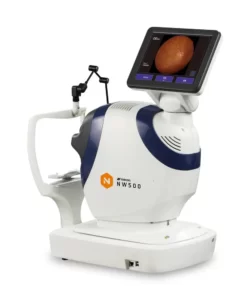
If your goal is to streamline retinal imaging while maintaining strong diagnostic quality, the Topcon NW500 is a smart, practical investment.
It won’t replace advanced multimodal systems—but it will improve your daily workflow, reduce staff burden, and keep your clinic running efficiently.
8. NIDEK Retina Scan Duo 2 ($3,333.00)
The NIDEK Retina Scan Duo 2 is a dual-imaging system for diagnosing macular and disc pathologies. It offers strong diagnostic flexibility for practices serving a patient base with diverse needs.
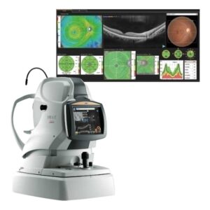
If you’re looking to streamline your imaging while maintaining strong diagnostic performance, the NIDEK Retina Scan Duo 2 is a very practical investment.
It simplifies your workflow, improves consistency across staff, and strengthens your ability to diagnose and monitor retinal conditions—all in one system.
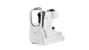
9. Canon CR-2 Plus AF
The Canon CR-2 Plus AF is a reliable digital, non-mydriatic retinal camera that provides both color and Fundus Autofluorescence (FAF). It offers consistent performance for practices of any size.
If you’re running a modern optometry or ophthalmology clinic and want a dependable, automated retinal camera with advanced capabilities, the Canon CR-2 Plus AF is a strong choice.
It improves workflow, enhances diagnostic confidence, and keeps your clinic running efficiently.
10. Cellview WRI-1
The Cellview WRI-1 is a budget-friendly widefield system that offers up to 133° in a single capture or up to 200° auto-stitched image. It’s a good entry-level option for new practices looking to get set up with the latest technology.
If you’re looking to upgrade from a traditional fundus camera and want wider retinal visibility, better screening, and improved workflow, the Cellview WRI-1 is a very practical choice.
It’s not trying to compete as the most premium system—it’s designed to be usable, efficient, and clinically meaningful.
Quick Comparison Table
|
Machine Name |
Type |
Field of View |
Key Strength |
Ideal Practice Type |
|
Optos Daytona |
UWF |
200° |
Fast scan time |
High-volume practices |
|
Oculus Pentacam |
Anterior segment analysis |
N/A |
Suitable for screening and refractive surgery |
Cornea-focused practices |
|
ZEISS CLARUS 700 |
UWF |
200° |
High-resolution imaging |
Practices conducting advanced diagnostics |
|
ZEISS CLARUS 500 |
UWF |
133º Widefield (one image) or 200º Ultra-widefield (two images) |
True color imaging |
Mid-sized practices |
|
Topcon Maestro2 |
OCT/fundus camera combination |
12mm x 9mm widefield |
Automation |
Small- to mid-sized practices |
|
iCare DRSplus |
Portable fundus camera |
80° |
Ease of use |
Practices doing routine screenings |
|
Topcon NW500 |
Fundus camera |
50° |
High-efficiency operation |
High-volume practices |
|
NIDEK Retina Scan Duo 2 |
Dual imaging OCT system |
12 x 9 mm widefield |
Scans both retina and macula |
Large practices |
|
Canon CR-2 Plus AF |
Non-mydriatic fundus camera |
45° |
No need for dilation |
High-volume practices |
|
Cellview WRI-1 |
Widefield |
133°(single-capture) 200° (auto-stitched image) |
Low-cost widefield system |
New practices |
Pros and Cons — A Realistic Look
There are notable advantages to UWF retinal imaging systems, including:
- Early disease detection
- Better documentation
- Improved patient communication
However, there are operational disadvantages that may affect adoptions, including:
- High upfront investment
- Staff training needed
- Maintenance costs
If you are unsure whether the benefits of an upgraded system are right for your practice, talk to one of the experts at Shape Ophthalmics. Our team can help you choose a system that improves practice efficiency and clinical outcomes while staying within your budget.
Final Thoughts
As you consider the right type of retinal imaging, it’s important to match the technology to your practice. If you have a high-volume clinic, you may benefit from an efficient ultra-widefield system. Multi-specialty clinics may need an OCT combo system. If you are a smaller practice, your team may work best with a fundus camera system.
The right machine is about more than just imaging. It can improve diagnosis, workflow, and patient trust.
FAQs
What are retinal imaging machines used for?
Retinal imaging machines capture detailed images of the retina. These images can reveal changes to the eye that may indicate eye disease such as glaucoma, macular degeneration, or diabetic retinopathy. Routine retinal imaging can identify issues before symptoms arise, leaving early treatment to preserve vision.
Do retinal cameras require dilation?
Some retina cameras require dilation. Others, such as ultra-widefield imaging systems, do not require pupil dilation.
What is ultra-widefield retinal imaging?
Ultra-widefield retinal imaging uses advanced laser technology to scan the retina and produce a detailed image. This technique captures more of the retina in a single image, enabling doctors to examine up to 85% of the retina, compared to 45% with traditional imaging.
How much does a retinal imaging machine cost?
The costs vary depending on the type of machine and the technology used. Small fundus cameras may cost only a few thousand dollars, while UWF systems may cost $20,000 or more.
Which system is best for small clinics?
Small clinics can often benefit from a standard fundus camera rather than a high-cost UWF system. These reliable systems are ideal for routine screenings in patients with low- to average retinal disease risks.